The following images are available for download. Unless otherwise noted, please credit Microbicide Trials Network, University of Pittsburgh and Magee-Womens Research Institute. If you have questions about these images or would like additional caption information, please contact Lisa Rossi at rossil@upmc.edu.
Microbicides
|
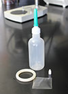
different formulations
|
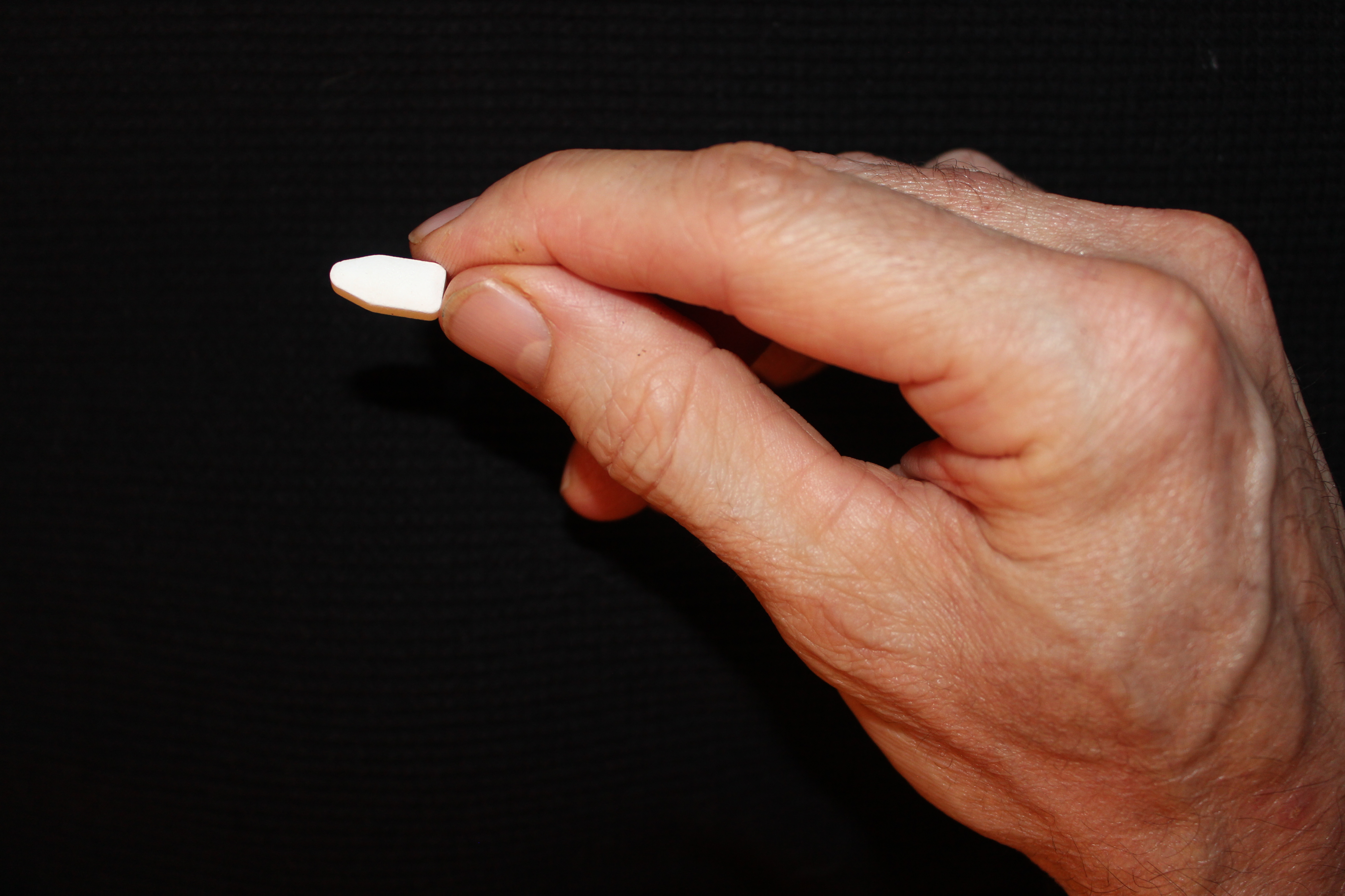
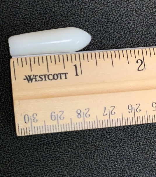
vaginal ring |
vaginal ring |
placebo insert |
placebo douche |


|
placebo suppository |
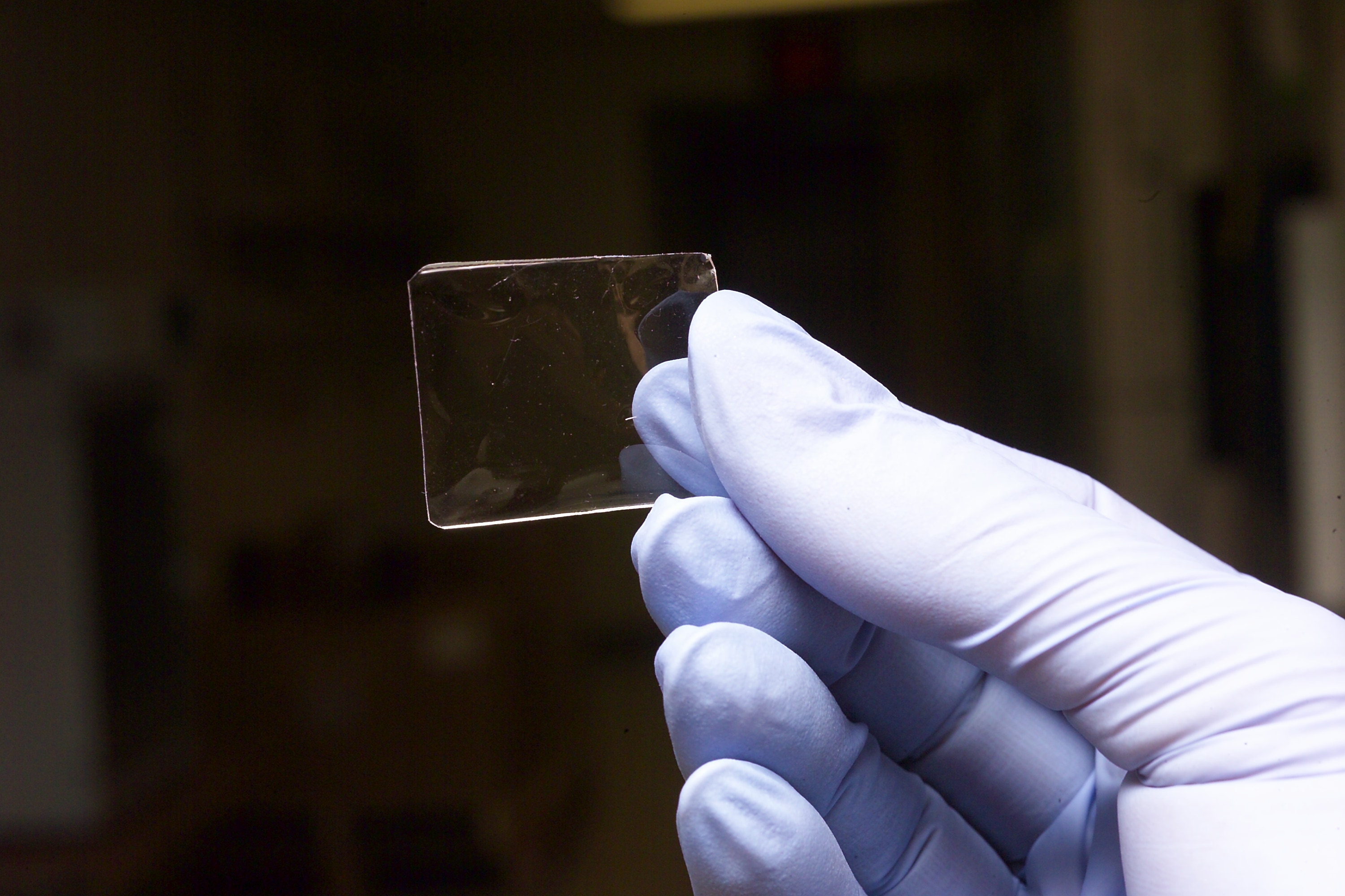
microbicide film |
microbicide film |
study gel and applicator
|
|
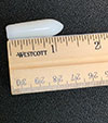


Microbicides can be formulated in different ways.
Research
|
Research 1 |
Research 2 |


Using a unique tissue explant model that very closely mimics how HIV infects cells of the cervix or rectum, MTN researchers are able to test different products for their safety and effectiveness.
|
Research 3 |
Research 4 |
Research 5 |


MTN researchers perform studies that assess the stability of drug compounds in different formulations of microbicides.
|
Research 6
|

MTN researchers look closely for any effects that microbicides may have on the normal population of vaginal microflora, studies that are important for assessing the safety of different products.
People
|
Sharon Hillier |
Elizabeth Brown Principal Investigator Statistical Data & Management Center Credit: FHCRC |
Connie Celum Principal Investigator Leadership and Operations Center UW Support Core Credit: University of Washington |
|
Kristine Torjesen Principal Investigator Leadership and Operations Center FHI 360 Support Core
|
John Mellors Principal Investigator Laboratory Center University of Pittsburgh |
Lisa Rohan Principal Investigator Laboratory Center University of Pittsburgh
|


Graphics and Illustrations
|
|

HIV infects T cells, one of several immune system cells, which have a specific molecule on its surface called a CD4 receptor. The receptor serves as a docking station where HIV sits before invading the cell. T cells are not found on the surface of the vagina, but below the epithelium, these and other target cells are found in abundance. How HIV reaches these cells is not certain. Perhaps HIV hitches a ride with dendritic cells that straddle the two layers, having conveniently been captured by these cells as an “invader” to be turned over to T cells and other immune cells that would otherwise orchestrate an attack. Or maybe the virus uses more direct routes through breaks in the tissue caused by local trauma and/or a sexually transmitted infection.